
Figure 4.
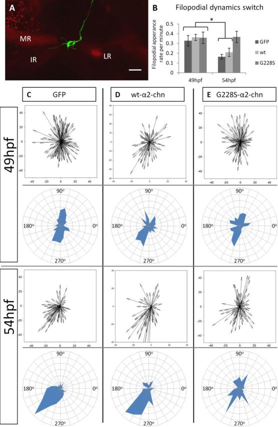
Filopodial dynamics during the 52 hpf transition. A, Illustrative image of a single GFP-expressing neuron within the OMN contacting the MR/IR progenitors in the alpha-actin-RFP line. B, Filopodial appearance rate in single neurons expressing either GFP, WT-α2-chn or the G228S-α2-chn mutant form both before and after the 52 hpf transition. *p <0.001. n = 9, n = 4, and n = 5 neurons, respectively, for 49 hpf. n = 10, n = 8, and n = 6 neurons, respectively, for 54 hpf. There is no significant difference between the rate of filopodial appearance in G228S-α2-chn-expressing neurons compared with GFP-expressing neurons either at 49 or 54 hpf (p = 0.869). C–E, Polar plots and radar plots showing the filopodial distribution before and after the 52 hpf transition in neurons expressing constructs as indicated. C, GFP-expressing neurons; the distribution at 49 and 54 hpf is different (p < 0.001). n = 173 and n = 93 filopodia, respectively. D, WT-α2-chn-expressing neurons; the distribution is different at 49 and 54 hpf (p = 0.037). n = 89 and n = 70 filopodia, respectively. The filopodial distributions for GFP and WT-α2-chn-expressing neurons are not significantly different either before or after (p = 0.417 and p = 0.445, respectively). E, G228S-α2-chn-expressing neurons; the distributions are similar (p = 0.985). n = 128 and n = 89 filopodia for before and after, respectively. The filopodial distributions for G228S-α2-chn-expressing neurons at 49 hpf or 54 hpf are not significantly different from those of either GFP or WT-α2-chn-expressing neurons at 49 hpf (p = 0.513 and p = 0.718, respectively).