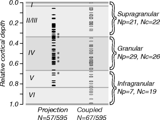
Figure 2.
Location and proportion of V2-connected neurons. Relative cortical depth of projection (N = 57) and coupled (N = 67) neurons whose visual selectivity we characterized (595 neurons recorded in total). Depth was computed as a fraction of the distance along recording tracks (from brain surface to white matter); laminar boundaries were drawn based on measurements of macaque Nissl-stained tissue sections (Tyler et al., 1998). Projection neurons (∼10% of all neurons recorded) were concentrated in cortical layers 2/3 and 4; coupled neurons were found in all layers. Asterisks show the locations of six doubly connected neurons; these were concentrated in the middle layers. Numbers of projection and coupled neurons in different laminar compartments (supragranular, granular, and infragranular) are listed (right; Np and Nc, respectively).

