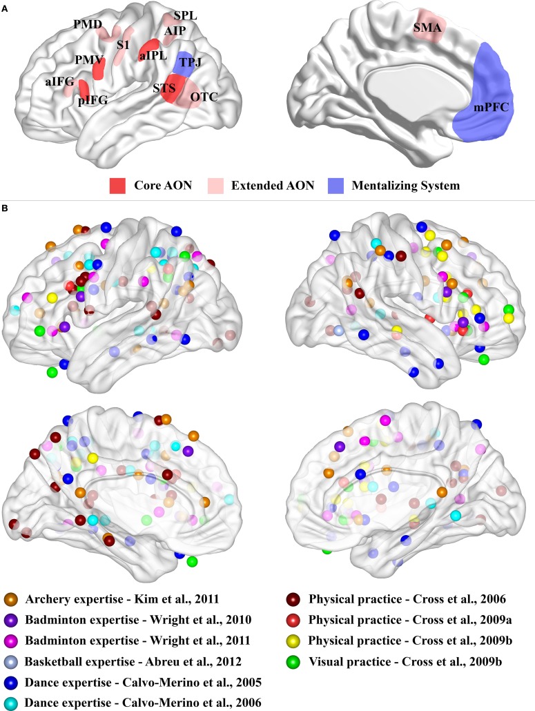
Figure 1.
(A) Schematic representation of the core AON, extended AON and mentalizing system. Three-dimensional representation of lateral and medial brain surface. The regions assigned to the “core” AON, “extended” AON and the “mentalizing” system are depicted. In red, the core AON is presented comprising: the PMV/pIFG complex, the aIPL and the STS. In pale red, the “extended” AON is presented comprising: the anterior part of the inferior frontal gyrus, (aIFG), the dorsal premotor cortex (PMD), the supplementary motor area (SMA), the superior parietal lobule (SPL), the anterior intraparietal sulcus (AIP), the somatosensory cortex (S1) and the occipito-temporal cortex (OTC), including also STS. The mentalizing system (blue) is assumed to consist of the medial prefrontal cortex (mPFC) and the temporo-parietal junction (TPJ). Note that the extension of these networks is not representative of their real dimension or functional significance. (B) Expertise effects. Three-dimensional representation of lateral and medial brain surface with location of peaks for the comparisons of interest superimposed. For Kim et al. (2011), we considered the comparison between the two groups (Table 2). For Wright et al. (2010), we considered results from ROI analysis (Table 2). For Wright et al. (2011), we plotted results for normal video (Table 2). For Abreu et al. (2012), we used the peak of the significant cluster within the temporal lobe in the group comparison (page 1649 of the manuscript). For Calvo-Merino et al. (2005), we plotted the reported interaction (see Table 1). For Calvo-Merino et al. (2006), we plotted the results from Table S2. For Cross et al. (2006), we considered the main effect of the contrast of interest (Table 2). For Cross et al. (2009a), we reported the contrast for physical training (Danced > Untrained) (Table 1). For Cross et al. (2009b), we considered the physical training results (Table 1) and the observational training results (Table 1). We excluded the peaks located within the cerebellum.

