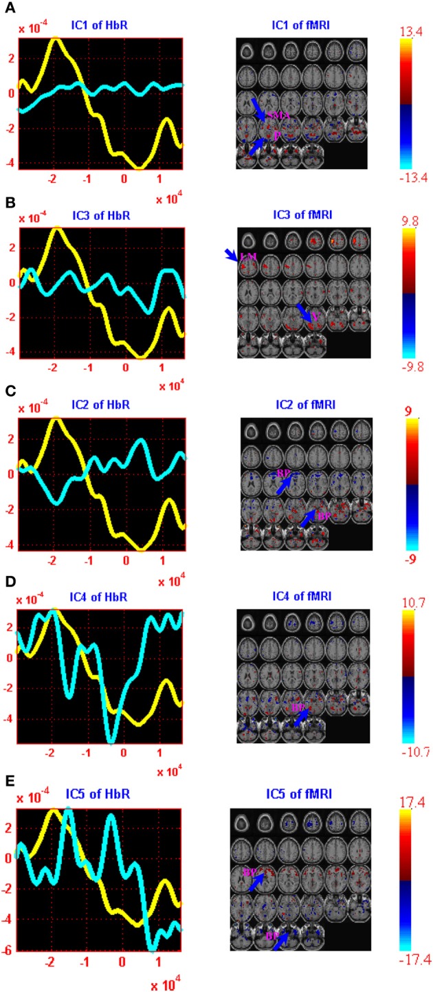
Figure 3.

jICA decomposition of ΔHbR and fMRI joint data for right finger tapping tasks: two components were found to be significantly correlated with right finger tapping task (ICs 1 and 3) while three were well-correlated with the physiology and body movement noise (ICs 2, 4, and 5). Each of these components is shown in a separate panel in the figure: IC1 (A), IC3 (B), IC2 (C), IC4 (D) and IC5 (E). The fMRI maps are thresholded at |Z| > 1.5 for display purposes. The averaged event-related HbR time course is shown in yellow (same for all figures) and the ΔHbR component is plotted in cyan. Positive (orange) and negative (blue) Z-values are shown in the image. The axes (bottom) illustrate the time scale, in ms, whereas the scale (left) records chromophore concentrations in μM for the figures on the left column. The scale for the figures on the right column shows the bold signal intensity. LM, Left primary motor cortex; SMA, Supplementary motor area; P, Parietal cortex; V, Visual cortex; BP, Body movement/physiology noise.
