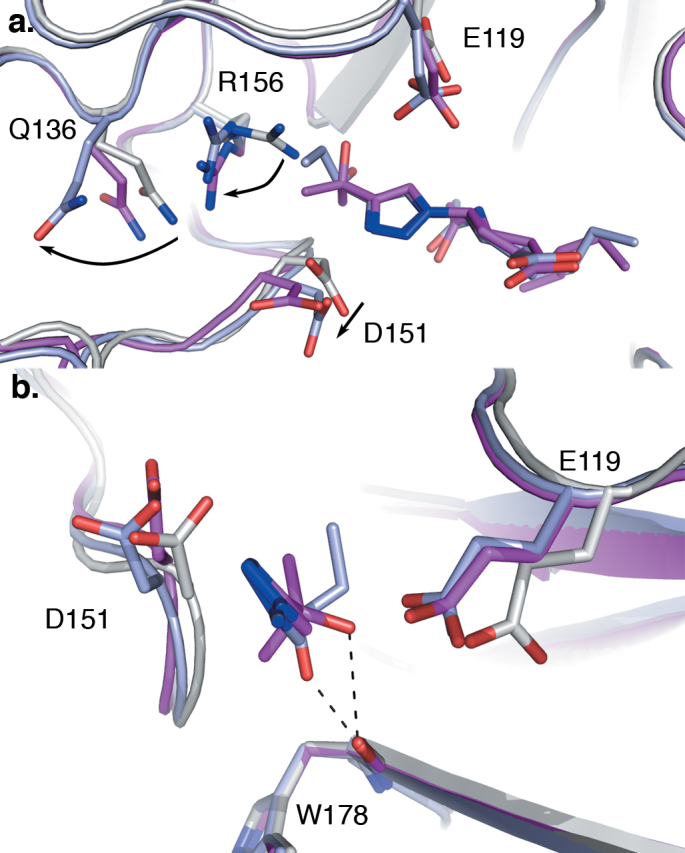
Figure 5. Interactions of 7 and 8 with the 150-cavity of the N8 active site.

Superimposition of N8:7 (pale blue) and N8:8 (magenta) with apo-N8 (white, 2HT5), with the atoms of ligands and relevant side chains shown as sticks. (a) Rearrangements within the 150-cavity of N8:7 and N8:8 in response to ligand binding (side chain movements indicated with arrows). Insertion of the triazole-hydroxyl moiety into the 150-cavity results in a repositioning of the guanidino group of R156, and resultant displacement of the Q136 side chain. Additionally the carboxylic acid of D151 moves 1.5 Å out of the active site to accommodate the triazole group. (b) View from the enzyme active site towards the 150-cavity, showing the interactions between the ligand triazole-hydroxyl moieties and residues within the active site (both shown as sticks, the cyclohexene ring and associated groups have been removed to improve visibility). Hydrogen bonding interactions formed between the triazole-hydroxyl moieties of 7 and 8, and W178 main-chain carbonyl are indicated with dashed lines.
