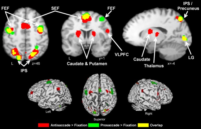
Figure 1.
Activation likelihood maps for antisaccade > fixation (red) and prosaccade > fixation (green). Regions where the contrasts overlap are shown in yellow. Abbreviations: FEF, frontal eye field; SEF, supplementary eye field; SPL, superior parietal lobule; IPS, intraparietal sulcus; IFG, inferior frontal gyrus; LG, lingual gyrus.

