
Figure 4.
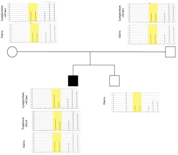
Pyrosequencing analysis of the DISP1 mosaic mutation [c.4412C>G (p.Ala1471Gly)] in Patient 1 and his family members. The mutated base (C>G) is highlighted in yellow. Quantitative analysis identifies that the mutation is present in 43%, 12%, 4.5% of the patient’s samples from lymphoblastoid cell line, peripheral blood cells, and saliva, respectively.