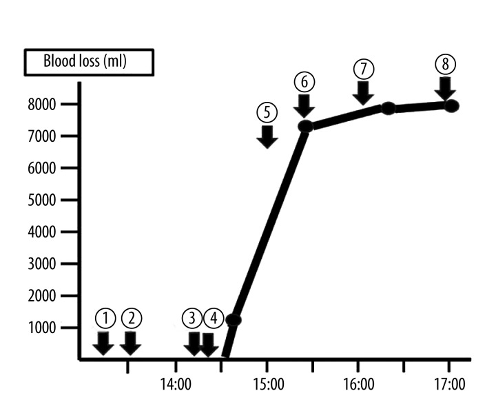
Figure 1.
Progress in the operation and blood losses. 1. The patient entered a operation room. 2. The preoperative occlusion balloons were placed in the bilateral internal iliac arteries by a radiologist. 3. We started the cesarean section. 4. 14:25 A male baby was born. The balloons in the bilateral internal iliac arteries were inflated, and we started the hysterectomy. 5. 15:00 A radiologist immediately transferred the balloon from the internal iliac artery to the common iliac artery. 6. 15:15 We restarted the hysterectomy. 7. We removed the uterus, and the balloons were deflated. 8. The operation finished.

