Figure 7.

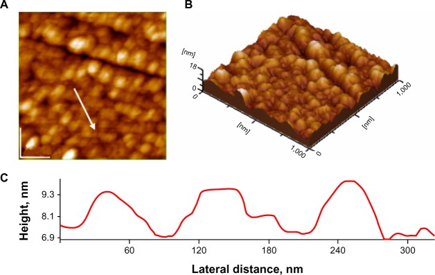
Atomic force microscopy images silver nanoparticles on glass in presence of 10−2 M dibucaine solution; scanned area 1 μm × 1 μm: (A) 2-D topography; (B) 3-D topography; (C) profile of the cross-section along the arrow in panel A.

Atomic force microscopy images silver nanoparticles on glass in presence of 10−2 M dibucaine solution; scanned area 1 μm × 1 μm: (A) 2-D topography; (B) 3-D topography; (C) profile of the cross-section along the arrow in panel A.