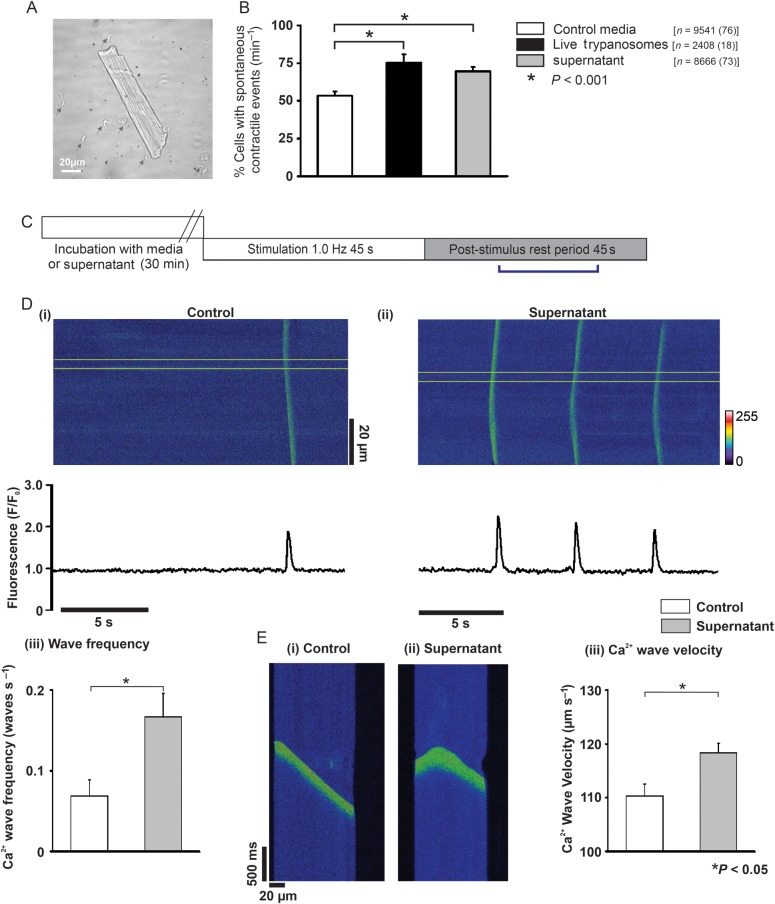
Figure 1.
(A) Isolated rat cardiomyocyte incubated with Trypanosoma brucei (grey arrowheads). (B) % cardiomyocytes with spontaneous contractile events (n = cardiomyocytes with number of hearts in parentheses). (C) Protocol used in separate confocal experiments. (D) Upper (i and ii) confocal line-scan images of cardiomyocytes from bracketed region in (C). Lower (i and ii) respective line profile trace taken from a 20 pixel region (denoted by yellow lines in upper image). (iii) Mean Ca2+ wave frequency; media (n = 18) and supernatant (n = 21). (Ei and ii) Individual Ca2+ waves, (iii) mean Ca2+ wave velocity in media (n = 53 waves from 18 cells) and supernatant (n = 158 waves from 21 cells).

