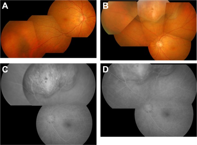
Figure 1.

Findings at presentation. (A) Fundus photograph of right eye. An orange-yellow mass is shown at the inferotemporal side. (B) Fundus photograph of left eye. A yellow-white mass is shown at the superior side. (C) Early frame and (D) late frame of fluorescein angiogram of left eye. Note hyperfluorescence in the tumor region from the early phase to late phase.
