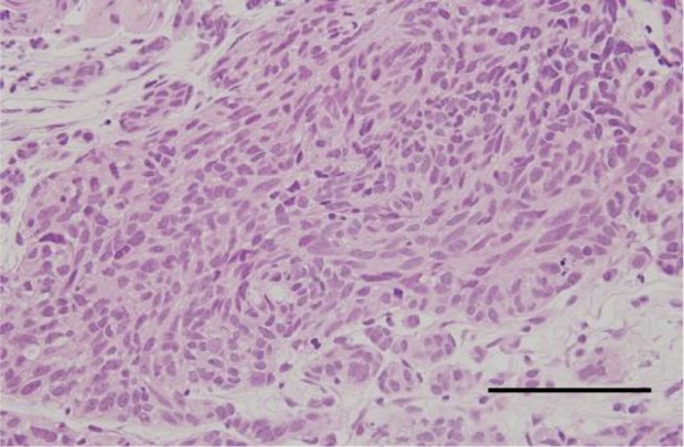
Figure 2.

Histopathology of the tumor from a breast biopsy. Foci of oval and spindle-shaped cells are shown in alveolar and palisading arrangement.
Notes: Hematoxylin and eosin staining; scale bar =100 μm.

Histopathology of the tumor from a breast biopsy. Foci of oval and spindle-shaped cells are shown in alveolar and palisading arrangement.
Notes: Hematoxylin and eosin staining; scale bar =100 μm.