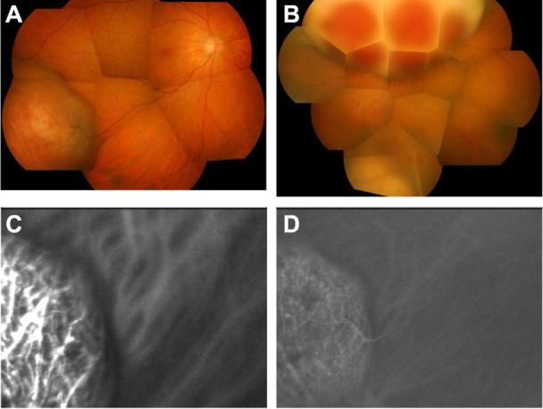
Figure 3.

Findings at 15 months after presentation. (A) Fundus photographs of right eye and (B) left eye. The choroidal tumors are shown. The tumors have apparently enlarged in size compared with size at presentation. (C) Early frame and (D) late frame of indocyanine green angiogram of right eye. Choroidal vessels inside the tumor are stained from the early phase, with a mixture of hyperfluorescence and hypofluorescence in the late phase.
