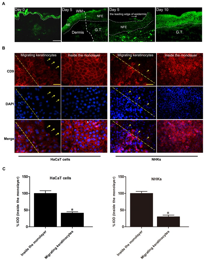
Figure 1. Downregulation of CD9 in keratinocytes at wound margin in vivo and in vitro.
(A) Immunofluorescence staining of CD9 in normal skin (Day 0) and wounded skin (Day 5 and Day 10) sections obtained from wild-type mice showing downregulation of CD9 in migrating epidermis during wound re-epithelialization. Wounds were fully re-epithelialized on day 10. Scal bar: 100μm. GT, granulationtissue; WM, wound margin; NE, normal epidermis; NFE, newly formed epidermis. Narrow-dotted line: interface between epidermis and dermis or leading edge of migrating epidermis (Day 5, right panel). Wide-dotted line: wound margin. (B) Immunofluorescence analysis of CD9 in HaCaT cells and normal human keratinocytes (NHKs) wounded using a micropipette tip (red labelling, 18 hours after wounding). Nuclei were stained with DAPI (blue). In the upper panel, the dotted line corresponds to the wound incision and the arrows indicate the direction of migrating keratinocytes. The lower panel shows images depicting the expression of CD9 in keratinocytes far from the wounded area of the cell monolayer. Scalebar: 50 μm. (C) Graphs to the right represent the corresponding % Integral optical density (IOD) of CD9 (red) (values were normalized as percentage after comparison with the keratinocytes inside the monolayer, which were set to 100%; n = 3). Results showed that CD9 expression in migrating keratinocytes was significantly downregulated. *P<0.05 vs. Inside the monolayer.

