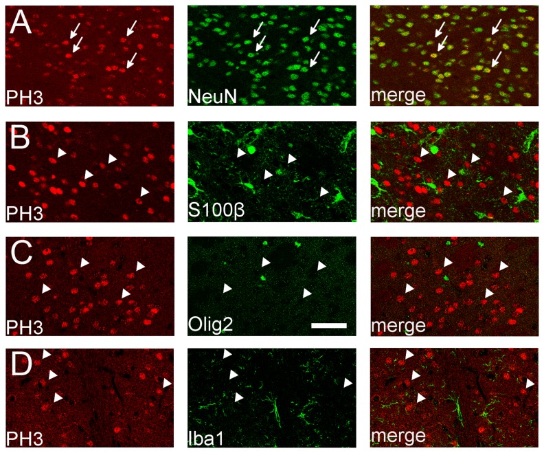
Figure 2. Identification of PH3+ cells in the CPu of the SE brain at 1 h.
Identity of PH3+ cells (red) was determined by double immunostaining with cell type specific markers (green). Virtually all the PH3+ cells are NeuN+ mature neurons (A), not S100β+ astrocytes (B), Olig2+ oligodendrocytes/oligodendrocyte precursors (C) or Iba1+ microglia (D). Arrows indicate PH3+/NeuN+ (double-labeled) cells and arrowheads indicate PH3+ (single-labeled) cells. Single optical confocal microscopy images are shown. Scale bar = 50 μm.

