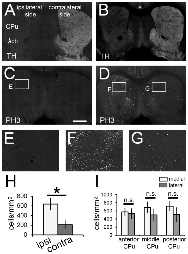
Figure 6. Dopaminergic input from the substantia nigra affects H3 phosphorylation in the CPu of the pilocarpine-induced SE brain.
Unilaterally 6-hydroxydopamine (6-OHDA)-injected mice were subjected to SE induction. Double immunostaining with anti-tyrosine hydroxylase (TH) and anti-PH3 antibodies was performed on the vehicle-administered control brain section (A, C and E) and the pilocarpine-induced SE brain section (B, D, F and G). TH immunoreactivity in the CPu and Acb is reduced significantly in the 6-OHDA injection side (A and B). H3 phosphorylation is significantly increased in the SE brain, especially in the ipsilateral side (D, F and G). Stacked epifluorescence microscopy images are shown. High magnification images of the boxed areas are shown in each image. (H) Density of PH3+ neurons in the CPu is significantly higher in the ipsilateral side than in the contralateral side of the pilocarpine-induced SE brain. White bar, ipsilateral side; gray bar, contralateral side. (I) Density gradient of PH3+ neuron in the CPu is lost in the 6-OHDA-injected side. White bar, medial part of the CPu; gray bar, lateral part of the CPu. Five animals were analyzed (n = 5). Two-tailed Welch’s t-test. *p < 0.05. Scale bar = 600 μm for A-D and 150 μm for E-G.

