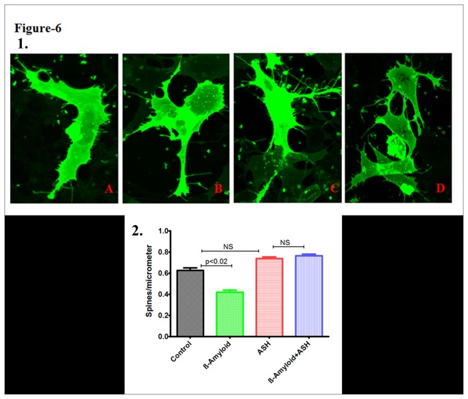
Figure 6. Confocal Images of DIL stained SK-N-MC cells showing the effect of β-amyloid1-42, Ashwagandha and Ashwagandha plus β-amyloid1-42.
1. A. Control, B. β- Amyloid 1-42 treated, C. Ashwagandha treated and D. Ashwagandha plus β- Amyloid1-42 treated. 2. Quantitative analysis showing the decreased spine density in β- Amyloid1-42 treated SK-N-MC cell line and its reversal by Ashwagandha (ASH). SK-N-MC cells were grown onto the glass coverslips, DIL stained and observed under confocal microscope. Randomly selected pictures in each group of the cells were captured in confocal microscope. Image J software was used to analyze the spine density, spine area, spine length and number of spines.

