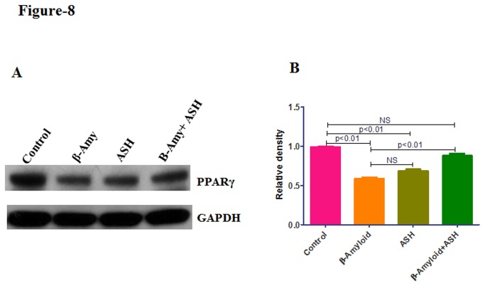
Figure 8. Western blotting analysis showing the decreased PPARγ protein levels in β-amyloid treated and its reversal by Ashwagandha in SK-N-MC neuronal cells.
(A) Cell lysates were separated in 4% to 15% linear gradient SDS-PAGE gels and were probed against the respective antibodies. GAPDH was used as the loading control. (B) Quantitative analysis showing the decreased PPARγ protein levels in β- Amyloid1-42 treated cultures. ASH - Ashwagandha; β-amy - β-amyloid. The gel shown is a representative for three experiments.

