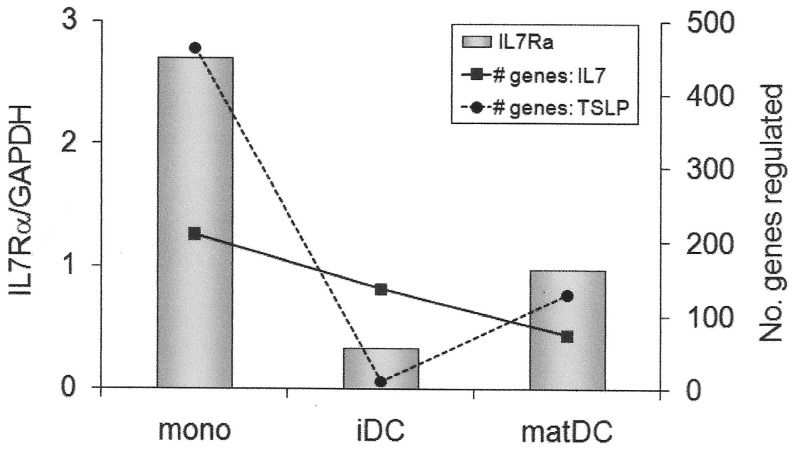
Figure 5. Magnitude of response to IL-7 or TSLP is dependent on IL7Rα expression in IFNβ-treated myeloid cell subsets.
Freshly purified monocytes (mono), in vitro cultured immature (IL-4, GM.CSF; iDC) and maturing monocyte-derived dendritic cells (IL-4, GM.CSF, LPS; matDC) from a heterozygous Hap2/Hap4 individual were incubated with IFNβ (1000 IU/ml) for 24 h. Bars represent the level of expression of IL7Rα after 24 h of IFNβ stimulation (IL7Rα). IL-7 or TSLP (10 ng/ml) were added at this point, and gene expression measured 24 h later. Lines represent the number of genes up- or down-regulated at least 1.5-fold by IL-7 (# genes: IL7) or TSLP (# genes: TSLP) as assessed by microarray. (These data also presented as individual numbers of up- and down-regulated genes in Table 1).

