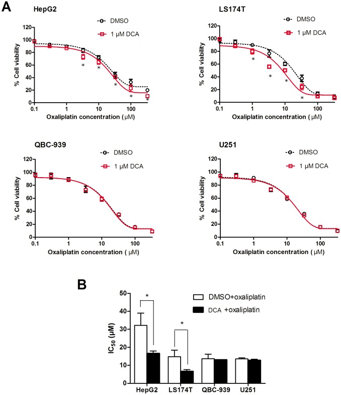
Figure 7. Demethylation of OCTN2 by DCA influenced cell viability to oxaliplatin.
Cancer cells were pre-treated with 1 μM DCA for 1 week, seeded in 96-well plates at 1×10⁴ cells/well in 100 μl DMEM and 10% FBS. After 8 h, the cells were exposed to oxaliplatin at various concentrations (0.1, 0.3, 1.0, 3.3, 10, 33, 100 or 330 μM) for 48 h in fresh culture medium. Cell viability was determined by a MTS cell proliferating assay kit. Each concentration was determined in three wells. Data presented represent the mean ± S.D. of three independent experiments. (A), the cell viability at each concentration was displayed. Significant differences from DMSO treatment are denoted with an asterisk (*, p<0.05). (B), IC50 values were calculated by SPSS 13.0 software. Data presented represent the mean ± S.D. of three independent experiments. Significant differences from non-treated cells are denoted with an asterisk (*, p<0.05).

