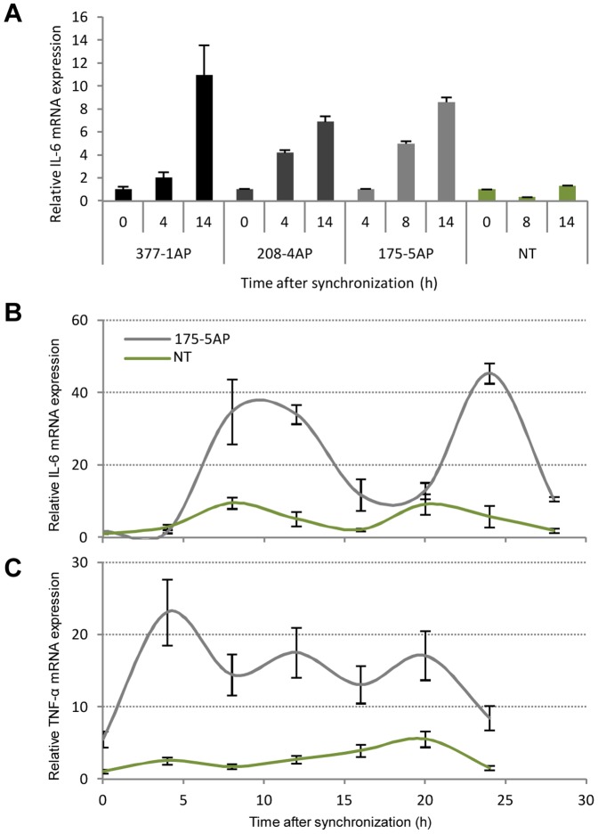
Figure 5. Induction of the proinflammatory cytokines IL-6 and TNF-α in fibroblast from the hCry1AP transgenic animals.
(A) Fibroblasts derived from skin-biopsies obtained from three transgenic minipigs (#377-1AP, #175-5AP, and #208-4AP) and one NT control (#321-1wt) were serum-shocked and allowed to recover. Subsequently, total RNA was extracted at three time points within 14 hours post-recovery. Quantitative RT-PCR was performed with exon-exon spanning primers targeting porcine IL-6 and normalized to endogenous ACTB. Values are relative to the first measurement time point. (B) Relative IL-6 RT-qPCR on total RNA extracted from serum-shocked fibroblasts originating from transgenic minipig #175-5AP and NT control pig #321-1wt as above. Cells were harvested every fourth hour through 24–28 hours. Values are relative to the first measurement time point (ZT 0). (C) Relative TNF-α mRNA expression in fibroblasts from transgenic minipig #175-5AP and NT control pig #321-1wt as above. All experiments are performed in triplicate and data are presented as mean values ± standard deviation.

