
Fig 4. Histamine H1 receptor in complex with doxepin (antihistamine drug).
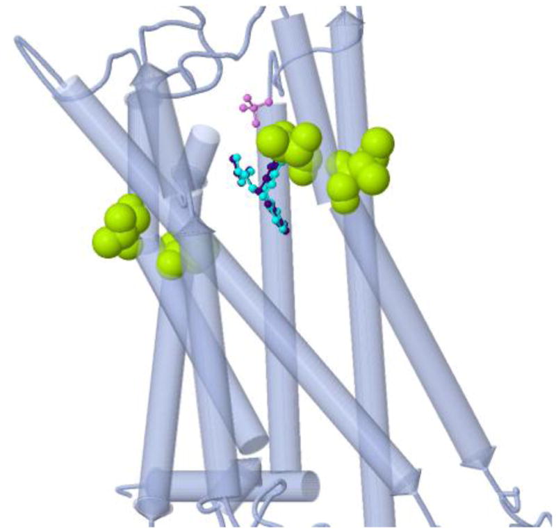
Nine variants from the ESP6500 (all with MAF < 1%) were mapped onto the PDB structure 3rze (Shimamura et al. 2011). Four variants that clustered proximal to the doxepin binding site are shown. Variant positions appear as green balls. Doxepin E-isomer is shown in dark blue. Doxepin Z-isomer is shown in light blue. Phosphate ion is shown in violet.