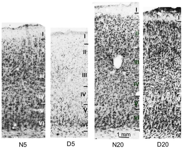
Figure 8.
Low magnification photomicrographs showing Abi-2 immunoreactivity in cat visual cortex. Coronal sections of Area 17 were processed for immunohistochemistry at the same time and in the same solutions in normal and dark reared cats at the peak (5 weeks: N5, D5) and nadir (20 weeks: N20, D20) of the critical period. Cortical layers are indicated and were determined from adjacent sections stained with cresyl violet. Calibration bar indicates magnification.

