
Figure 2. siORC1 impairs recovery of replication following HU and reduces HU-induced new origin firing in U2OS.
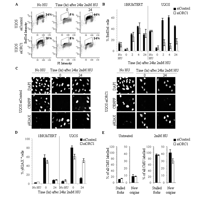
(A-B) 1BR3hTERT and U2OS cells were transfected with siRNA oligonucleotides (5 or 0.6 nM, respectively). 48 hours later, 2 mM HU was added for 24 hours and cells were grown for times shown. BrdU was added 30 minutes prior to processing by FACS. The fraction of replicating (BrdU+) cells was determined. (A) Representative FACS analysis. Boxed regions containing black data points indicate BrdU+ cells; numbers represent BrdU+ cell fraction. Supplementary Fig. S2A shows additional analyses. (B) Quantification from three experiments using 1BR3hTERT or U2OS cells. (C) Representative immunofluorescence images showing DAPI (DNA), CENPF (cell cycle phase) or γH2AX (DNA damage) in U20S cells treated as in (A-B). Representative merged channel images are shown in Supplementary Fig. S2B. (D) Nuclei containing bright γH2AX pan-nuclear staining were scored as γH2AX+. Figure shows the fraction of γH2AX+ cells. (E) 48 hours following transfection of U2OS cells with 0.6 nM siControl or siORC1, cells were pulse labelled with CldU, treated with 2 mM HU for 24 hours and released (or untreated), and pulse labelled with IdU for 1 hour. The number of structures representing fork stalling and new origin firing was normalised to the number of CldU+ replication tracks. The experimental design and representative images are shown in Supplementary Fig. S2C-D. Results represent the mean and SD of > 2 experiments (0mM HU n=2, 2mM HU n=3).