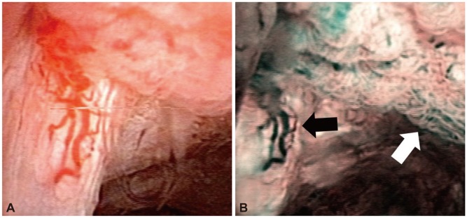
Fig. 2.

Comparison of conventional white light imaging and narrow band imaging in the case of recurred gallbladder cancer after cholecystectomy. (A) The white light imaging shows tumor vessels and a polypoid mucosal lesion. (B) The narrow band imaging shows papillary structures of mucosal surface (white arrow) and microvasculatures (black arrow) more definitely than white light imaging.
