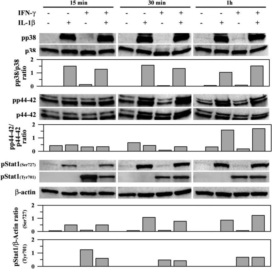
Fig. 7.
IL-1β- and IFN-γ-induced signaling pathways in human astrocytes. Human astrocyte cultures in 12-well plates (106 cells/well, 600 µl DMEM) were untreated (as control) or treated with IL-1β (10 ng/ml) ± IFN-γ(200 U/ml) for 15, 30 and 60 min before collecting cell lysates to be electrophoresed, transblotted to nitrocellulose membrane and probed for p38 or p44/42 MAPK, Stat1 or β-actin (as internal control). Densitometric measurement (normalized to total p38 or p44/42 or β-actin) of the bands was shown. Data presented are representative of three to five separate experiments using astrocytes derived from different brain tissue specimens. All targets of the same experiment were electrophoresed and analyzed in individual blot without re-probing.

