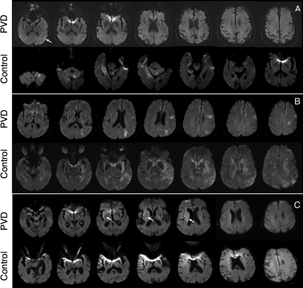
Figure 3.

Examples of diffusion weighted images (DWI) of case-control patient pairs at presentation of stroke. A) PVD: 80 year-old male with restricted diffusion within the left cortical parietal lobe (white arrows, hyperintense volume: 0.3852mL). Control: 83 year-old male with restricted diffusion in left cerebellum, right temporal and right occipital lobe (21.36mL). B) PVD: 70 year-old female with restricted diffusion in the posterior right MCA territory (121.9mL). Control: 70 year-old female with restricted diffusion in the left MCA, caudate and semiovale (201.1mL). C) PVD: 99 year-old female, restricted diffusion in the right parietal lobe (67.96mL). Control: 87 year-old female, restricted diffusion of the right frontal cortex, right insular cortex, and extending to anterolateral right frontal lobe cortex and corona radiata (12.09mL).
