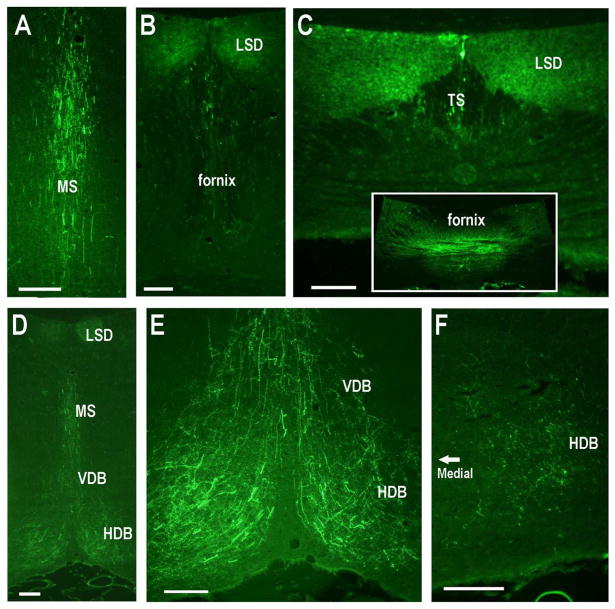
Fig. 12.
Subcortical projections of dorsal CA2 neurons following six-site injections (as shown in Fig. 10). A–C. Progressively further posterior sections showing GFP-positive fibers within the MS and then LSD, TS and fornix. D-F. GFP-positive fibers within diagonal bands of Broca. Scale bars equal 100 μm.

