
Fig 3.
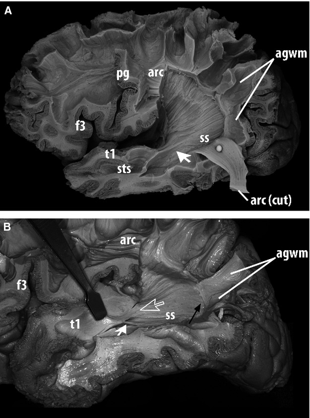
Lateral view of the dissection of the deep white matter of the left hemisphere of a human brain, a subsequent stage to Fig.2. After the arcuate fasciculus was caudally retracted, the plane between the arcuate fasciculus and deeper horizontal association fibers was the interface between the medial aspect of the arcuate fasciculus and the lateral layer of the sagittal stratum. (A) Horizontal fibers of this layer were followed throughout the dissection to assess whether they entered the superior temporal gyrus, which is exhibited in this specimen (arrow). (B) Another specimen, in which the parieto-temporal portion of the arcuate fasciculus was elevated and removed. Delicate groups of fibers from the most lateral layer of the stratum sagittale did not submerge in deep portions of the temporal lobe but remained relatively superficial on entering the superior temporal gyrus (large blank arrow). Additional groups of fibers entering the superior temporal gyrus at different points (small white arrow) could occasionally be exposed by gentle upward retraction of the temporal operculum. Although these fibers were encountered in the posterior and middle-thirds of T1/PT, the procedure failed to follow them for a long trajectory up to the temporal pole. (A,B) Posteriorly, these fibers stayed deep to the white matter of the angular gyrus without any fiber group shifting to its cortex: association fibers of the posterior bank of the angular gyrus were continuous with those of the arcuate fasciculus but discontinuous with fibers of the superficial layer of the stratum sagittale (black arrow). agwm, angular gyrus white matter; arc, arcuate fasciculus; f3, inferior frontal gyrus; ls, lateral sulcus; pg, pre-central gyrus; t1, superior temporal gyrus; sts, superior temporal sulcus, ss, stratum sagittale.