
Fig 4.
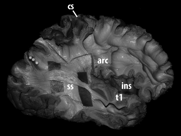
Lateral view of the dissection of the most external layer of the stratum sagittale of the right hemisphere of a human brain. The horizontal association fibers entering the region of the superior temporal gyrus were further dissected and exposed. In this process, most of the arcuate fasciculus was removed. Following those fibers posteriorly showed that they were directed to upper portions of the occipital lobe, next to the postero-superior border of the hemisphere and the parieto-occipital arcus. The level of the parieto-occipital sulcus was marked with three white pins. arc, arcuate; cs, central sulcus; ins, insula; t1, superior temporal gyrus; ss, stratum sagittale.