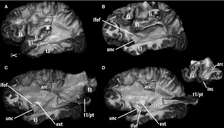
Fig 5.

Further dissection of the deep white matter along the stratum sagittale and the anterior temporal lobe in a human left hemisphere, lateral view. The fibers coming from the inferior fronto-occipital fasciculus need to be identified to assess whether they are distinguishable from those entering the superior temporal gyrus and to expose their relationships. (A) Fibers from the lateral layer of the stratum sagittale ran deep to the arcuate fasciculus and were dissected up to their posterior terminations in the occipital lobe. Anteriorly, they entered the superior temporal gyrus. In order to advance to the depth of the temporal lobe and insula, an incision was performed at the level of the superior temporal sulcus. (B) After the superior temporal gyrus including the planum temporale was detached from the deep temporal white matter, a plane was developed by passing through the insula, lateral to the external capsula. To dissect deeper structures, the superior temporal gyrus, the insular cortex and the inferior frontal gyrus were removed completely. The attachment of the superior temporal gyrus to the stratum sagittale was preserved. (C)The whole block comprising insular cortex and frontal and temporal opercula was fully elevated laterally, providing access to the structures in the external capsula. At this level, fiber dissection allowed identification of the uncinate fasciculus posterior to the limen insulae, and the inferior fronto-occipital fasciculus, located postero-superior to the uncinate fasciculus and running from the frontal lobe to the stratum sagittale. With this procedure, claustro-fugal fibers were also exposed in the postero-superior portion of the external capsula. (D) The insular cortex and the frontal operculum were detached from the temporal operculum. A divider was introduced deep to the fibers of the inferior fronto-occipital fasciculus, which were further dissected. This procedure showed that the iFOF was medial to fibers of the stratum sagittale reaching the superior temporal gyrus. Whereas the fibers of the stratum sagittale that form the iFOF inclinated medially and went deeper to reach the external capsula, those connecting the superior temporal gyrus did not incline and remained relatively superficial with a straighter trajectory. Also, in the stratum sagittale, iFOF fibers were more medial and slightly inferiorly situated. arc, arcuate fasciculus; ext, external capsula; f3, inferior frontal gyrus; ifof, inferior fronto-occipital fasciculus; ins, insula; ls, lateral sulcus; pt, planum temporale; t1, superior temporal gyrus; t3, inferior temporal gyrus; unc, uncinate fasciculus; ss, sagittal stratum.
