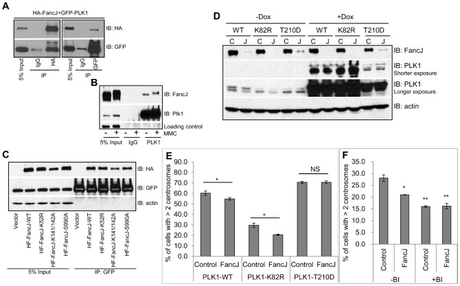
Fig. 5. FancJ binds PLK1 and promotes the activation of PLK1 during the MMC induced centrosome amplification.
(A) HA tagged FancJ and GFP tagged PLK1 were co-transfected into 293T cells. An equal amount of cell lysate was used for immunoprecipitation with either IgG or antibody against HA or GFP as indicated. Antibodies used for immunoblotting are indicated on the right. (B) 293T cells were either left untreated or treated with 0.5 µM of MMC for 24 hours. An equal amount of cell lysate was used for immunoprecipitation with either IgG or antibody against PLK1. Antibodies used for immunoblotting are indicated on the right. (C) Either vector or different HA tagged FancJ variants were co-transfected with GFP tagged PLK1 into 293T cells. An equal amount of cell lysate was used for immunoprecipitation with the antibody against GFP. Antibodies used for immunoblotting are indicated on the right. (D) Three different cell lines expressing PLK1 under control of a doxycycline (Dox)-inducible promoter were first transfected with either Control siRNA (C) or pooled siRNA against FancJ (J) and then split into two sets. Doxycycline was added to one set of the cells to induce the expression of different PLK1 variants (+Dox). 24 hours later, cells were collected and used for Western Blot analysis. Antibodies used for immunoblotting are indicated on the right. (E) The three Dox-regulated cell lines were first transfected with either Control siRNA (Control), or pooled siRNA against FancJ (FancJ), and then treated with doxycycline to induce the expression of different PLK1 variants. This was followed by treatment with 0.5 µM MMC for 72 hours. Cells were finally fixed in methanol and stained with antibody against γ-Tubulin. More than 300 cells were counted and the percentage of cells with more than two centrosomes was quantitated. (F) U2-OS cells were first transfected with either Control siRNA or siRNA against FancJ and then split into two sets. One set was treated with 0.5 µM MMC for 72 hours (−BI). In the second set, cells were first treated with 0.5 µM MMC for 12 hours and followed by the addition of 100 nM BI-2536 (+BI). Sixty hours after the addition of BI-2536, cells were finally fixed in methanol and stained with antibody against γ-Tubulin. More than 300 cells were counted and the percentage of cells with more than two centrosomes was quantitated. All error bars are standard deviation obtained from three different experiments. Standard two-sided t test, *P<0.05, **P<0.01, ***P<0.001, NS, no significance.

