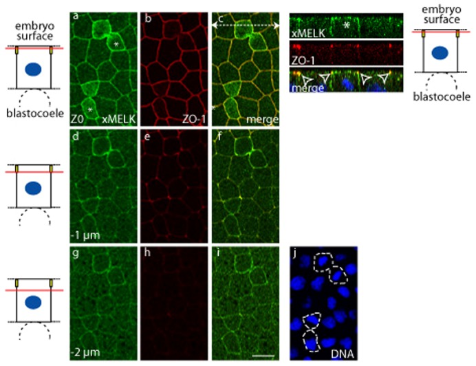
Fig. 3. In epithelial cells xMELK accumulates at the tight junctions.

Confocal microscopy of indirect immunofluorescence with anti-xMELK (green) and anti-ZO-1 (red) antibodies of epithelial cells from fixed albino embryos at gastrula stage. Three single optical sections spaced by 1 µm are shown. Asterisks indicate cytokinetic cells. Diagrams on the left: red lines mark the confocal planes relative to embryo surface and yellow rectangles symbolize tight junctions. Images were merged to visualize co-localization of xMELK with ZO-1 (merge, c,f,i). DNA is blue (j), dividing cells are indicated by dashed lines. White dashed arrow in panel c symbolizes the plane used for orthogonal projection of confocal planes shown on the right. Arrowheads point to the xMELK which co-localizes with ZO-1 at the tight junctions. Scale bar: 20 µm.
