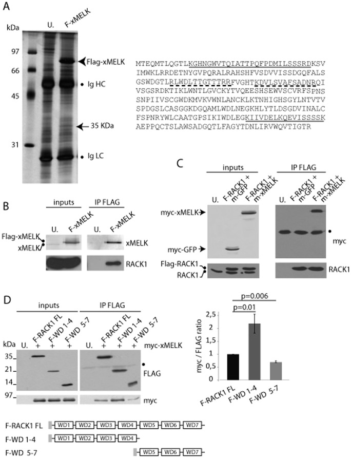
Fig. 6. xMELK and RACK1 are in the same complex.
(A) Identification of RACK1 as a potential xMELK partner. Proteins extracted from FLAG-xMELK expressing or uninjected control (U.) embryos were immunoprecipitated with anti-FLAG antibodies, separated by SDS-PAGE and silver stained. The 35 kDa band present in the FLAG-xMELK but not in the control immunoprecipitate was cut out from the gel and analyzed by mass spectrometry. Two peptides matching RACK1 protein sequence (underlined) were identified. Two additional peptides were identified in an independent experiment (dashed underline). Ig HC and Ig LC: immunoglobulins heavy and light chains, respectively. (B,C) Validation of xMELK and RACK1 interaction. (B) Proteins were extracted from FLAG-xMELK (F-MELK) expressing or uninjected (U.) embryos (inputs). Proteins were immunoprecipitated with anti-FLAG antibodies (IP FLAG) and Western blots were incubated with anti-xMELK and anti-RACK1 antibodies. (C) Protein extracts (inputs) were prepared from embryos co-expressing FLAG-RACK1 (F-RACK1) and myc-GFP (m-GFP), FLAG-RACK1 and myc-xMELK (m-MELK) or uninjected control embryos. Proteins were immunoprecipitated with anti-FLAG antibodies (IP FLAG) and Western blots were incubated with anti-myc and anti-RACK1 antibodies. (D) xMELK preferentially associates with RACK1 N-terminal domain. Protein extracts (inputs) were prepared from embryos co-expressing myc-xMELK with full length FLAG-RACK1 (F-RACK1 FL), FLAG-RACK1 WD1–4 (F-WD1–4), and FLAG-RACK1 WD5–7 (F-WD5–7) or uninjected (U.) embryos. Proteins were immunoprecipitated with anti-Flag antibodies (IP FLAG) and Western blots were incubated with anti-FLAG and anti-myc antibodies. The histogram on the right represents quantifications of the myc signal obtained in 3 independent immunoprecipitation experiments normalized with the corresponding FLAG signals (myc/FLAG ratio). Error bars denote s.e.m., a t-test was performed and p values are indicated above bars. Schematic representation of the RACK1 constructs is shown. The grey box indicates the FLAG tag.

