
Fig. 1. Impact of rbpj overexpression on dlc expression.
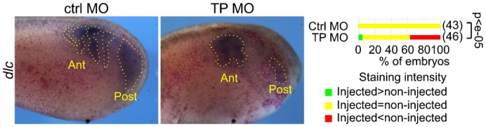
We injected nLacZ mRNA and control (left panel) or the target-protector (middle panel) morpholinos into one blastomere of two-cell Xenopus embryos and allowed the embryos to develop to the tailbud stage. We then stained them for β-galactosidase activity (red dots) and dlc by in situ hybridisation (ISH). The right panel shows the percentage of embryos with a staining intensity in the injected side above, equal to, and below (respectively green, yellow and red) that in the control side. We compared the distributions between these three classes by a chi-square test and we show the p-value. The photographs are lateral views of the injected sides, anterior left. The positions of the anterior PSM (Ant) and the posterior PSM (Post) are indicated.