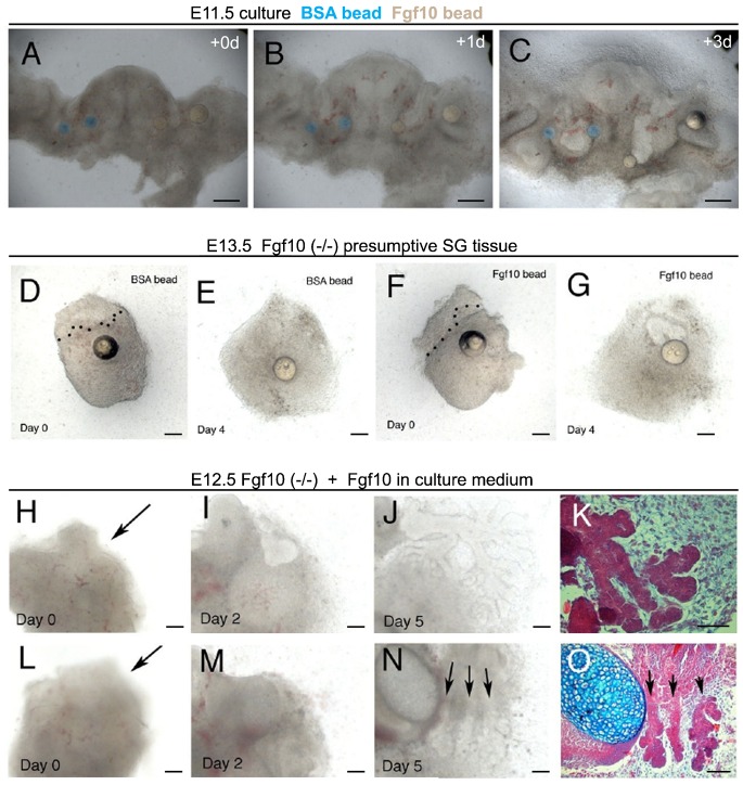
Fig. 5. Fgf10 leads to elongation of the salivary gland.
(A–C) Slice cultures of the developing mandible at E11.5. Frontal live section 200 µm. Tongue in the middle with developing salivary glands on either side. (A) Day 0. BSA control beads on LHS (blue). Fgf10 loaded Beads RHS (white). Submandibular glands at the initial bud stage. (B) Day 1. The epithelium on the RHS has turned towards the nearest Fgf10 bead, while the control side grows straight down. (C) Day 3. The epithelium on the RHS has elongated past the normal gland position pushing the bead downwards. Rescue of gland development. (D–O) Fgf10 mutant presumptive salivary gland tissue. (D,F) E13.0 gland with beads added. Day 0. Epithelium outlined by dotted lines. (E) Day 4. No growth of the epithelium in after addition of a PBS control bead. (G) Day 4. Growth of the epithelium towards the Fgf10 bead, and the start of branching morphogenesis. (H–O) E12.5 gland with Fgf10 added to the medium. (H,L) Day 0. Arrows point to gland epithelium arrested at the thickening stage. (I,M) Day 2. In panel I a clear bud can be observed extending into the mesenchyme that has condensed into a capsule. (J,N) Day 5. Branching glands are in evidence. In panel N three distinct glands have formed (arrows). (K,O) Histological sections of the cultures showing branching and the onset of lumen formation. Image shows merged image from two adjacent sections to show the whole gland. Scale bars: 250 µm (A–C), 100 µm (D–O).

