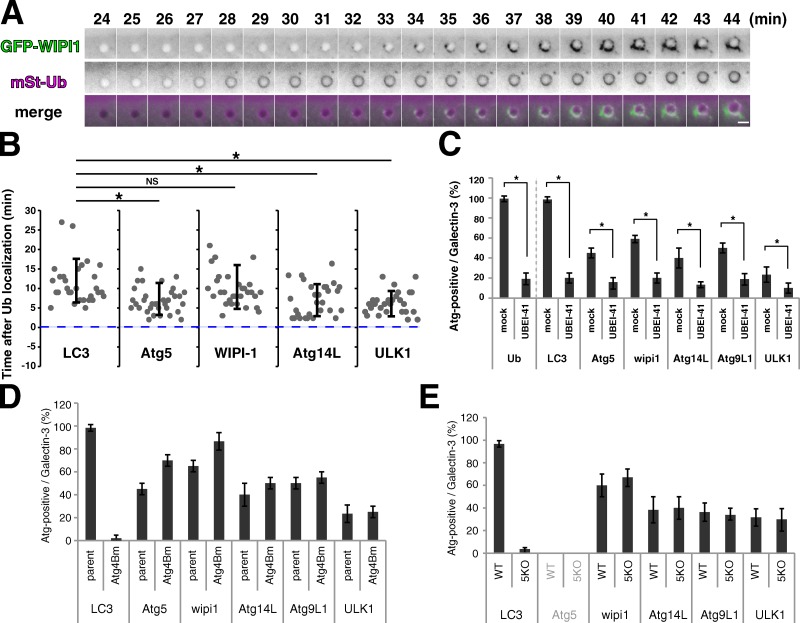
Figure 2.
Ubiquitination and recruitment of Atg proteins. (A and B) NIH3T3 cells stably expressing mStr-Ub and GFP-tagged LC3, Atg5, WIPI-1, Atg14L1, or ULK1 were transfected with Effectene-coated latex beads for 30 min. Then, live cells were observed at 1-min intervals by fluorescence microscopy. Bar, 3 µm. The time after Ub localization was measured for at least 30 cases for each combination. Each value represents the mean ± SD. Statistical analysis was performed by Student’s unpaired t test. *, P < 0.05; NS, not significant. (C) NIH3T3 cells stably expressing GFP-tagged Ub, LC3, Atg5, WIPI-1, Atg14L1, Atg9L1, or ULK1 were transfected with Effectene-coated beads for 3 h in the presence or absence (mock) of 30 µM UBEI-41 (a ubiquitin E1–specific inhibitor) and subjected to immunocytochemistry for galectin3. The percentages of GFP-positive per galectin3-positive beads were enumerated. At least 30 beads were counted (n = 3). The values are the mean ± SD. Statistical analysis was performed by Student’s unpaired t test. *, P < 0.05. (D and E) Parent NIH3T3 cells, Atg4B mutant overexpressing NIH3T3 cells (D), wild-type MEFs, and Atg5-KO MEFs (E) stably expressing GFP-tagged LC3, Atg5, WIPI-1, Atg14L1, Atg9L1, or ULK1 were transfected with Effectene-coated latex beads for 3 h and subjected to immunocytochemistry for galectin3. The percentages of Atg-positive per galectin3-positive beads were enumerated. At least 30 beads were counted (n = 3). The values are the mean ± SD.

