Figure 6.
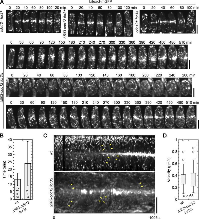
Medial actin filaments are predominant for contractile ring assembly. (A) Time courses of contractile ring assembly and constriction by tetrad fluorescence microscopy in cells expressing Lifeact-mGFP from the cross JW5463 × JW5481. The interval of the time-lapse movie is 10 min and time 0 is the time point just before actin filaments concentrate near the division site (except for the double mutant with no accumulation at the division site). Three classes of double mutant cells are shown: large clump at the division site (top); no specific structures (middle); and severely delayed ring assembly (bottom). (B) Timing of actin accumulation in a ring/meshwork (wt) or aster (Δ503-cdc12 for3Δ) after Rlc1-tdTomato node appearance in tetrad fluorescence microscopy. Many double mutants could not be included in this quantification because they did not attempt division during the imaging. Error bars show SDs. (C and D) Actin filament movements toward the division site. (C) Kymographs of wt and double mutant (Δ503-cdc12 for3Δ) cells expressing Lifeact-mGFP. De novo actin filament nucleation (dashed box, wt only) and cortical flow (examples are marked by pairs of yellow arrowheads) are evident in tetrad fluorescence microscopy from the same cross as B. (D) Box plots of velocity of actin movements toward the division site as shown in C. Bars, 5 µm.

