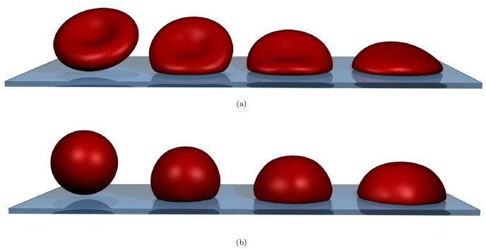
Figure 1. Simulated cell spreading of the red blood cell at three different time-points.
(a) binconcave RBC spreading. (b) “sphered” RBC spreading. From left to right: no contact at  , early contact at
  , approximately the cross-over between the two regimes at
  and the fully spread cell at
  . The biconcave RBC has approximately 40% less volume than the osmotically swollen spherical red blood cell. For movies corresponding to these snapshots, see supplementary Videos S1 and S2.

