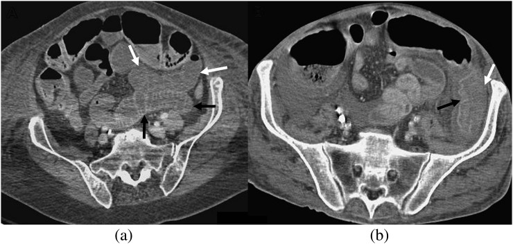
Figure 4.
Differential bowel wall enhancement. (a) A 79-year-old female patient, 1 day post coronary artery bypass graft. Adjacent loops of ileum show differential enhancement with hypo- (white arrows) and hyperenhancement (black arrow). (b) Axial CT in a different patient showing differential enhancement of the descending colon with hyperenhancement of the medial wall (black arrow) and reduced enhancement and oedema of the lateral wall (white arrow).

