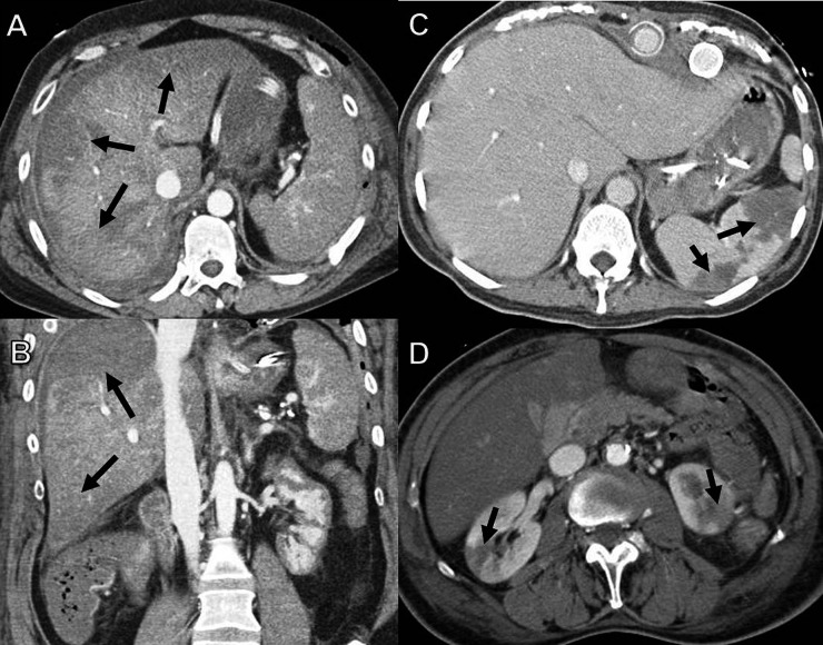
Figure 6.
Solid organ hypoperfusion/infarction. (a,b) Axial CT imaging (a) with coronal reformats (b) in a 41-year-old female patient, 4 days post coronary artery bypass graft surgery, shows peripheral wedge-shaped areas of hypoperfusion in the liver (arrows). (c) Axial CT in an 82-year-old female patient, 10 days post aortic valve replacement (AVR) with multiple splenic infarcts (arrows). (d) A 65-year-old male patient, 14 days post mitral and AVR surgery with bilateral renal infarcts (arrows).

