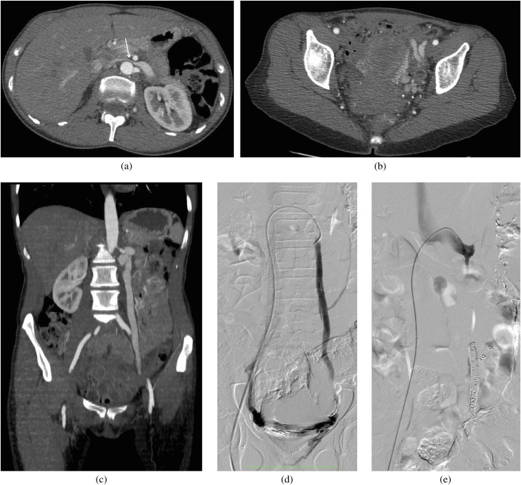
Figure 3.
A 25-year-old female who presented with pelvic pain and was also found to have haematuria. (a–c) A contrast-enhanced CT scan was performed to evaluate the cause of pain. An axial scan at the level of (a) the left renal vein (LRV) and (b) in the pelvis, and (c) a coronal scan through the gonadal vein demonstrate compression of the LRV (arrow) by the superior mesenteric artery anteriorly and the aorta posteriorly, multiple pelvic collateral vessels and an enlarged gonadal vein. (d–e) The patient later underwent a venogram to demonstrate the reflux and to coil embolise the gonadal vein. (d) Venogram via a right common femoral vein access and injection of the gonadal vein demonstrates an enlarged gonadal vein and cross pelvic collaterals with partial opacification of the right gonadal vein. (e) Venogram following coil embolisation of the left gonadal vein shows absence of reflux.

