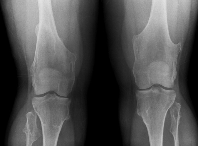
Figure 2.

Knee radiographs of a young patient with hereditary multiple exostoses showing multiple exostoses in the distal femur and proximal tibia and fibula, which typically point away from the joint. There is an associated widening of the diametaphysis resulting in an Erlenmeyer flask deformity.
