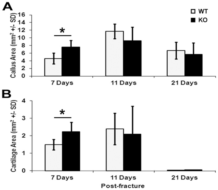
Figure 2. Quantification of callus and cartilage development during fracture healing.
Histomorphometric quantification of cartilage area (A) and fracture callus areas (B) derived from WT and Darc-KO mice at 7, 11 and 21 days post-fracture. We examined 5–9 animals/time point/strain of mice. *p<0.05 WT vs Darc-KO mice.

