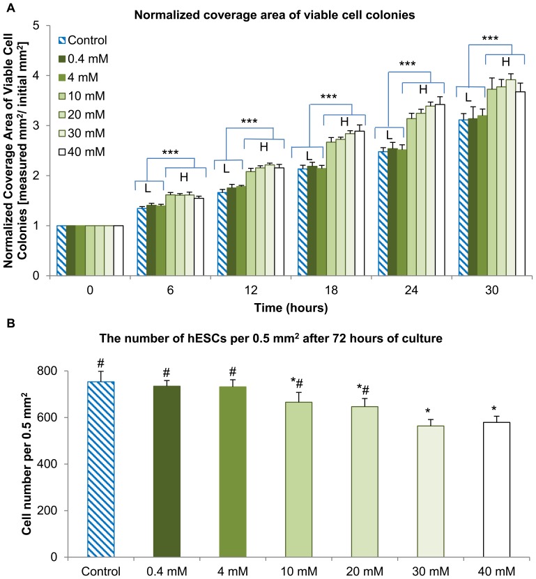
Figure 6. Coverage area of viable hESC colonies and the numbers of hESCs when cultured in the media supplemented with Mg ion dosages.
(A) The coverage area of viable hESC colonies after being normalized over the first time point at 0 hour in the cultures with respective supplemental Mg ion dosages (***p<0.05 when comparing L-group with the H-group). (B) The average numbers of hESCs per 0.5 mm2 after being cultured for 72 hours and immunostained with DAPI and pluripotency marker-SOX2 (*p<0.05 compared to the control and #p<0.05 compared to 40 mM). Although the normalized coverage area of viable hESC colonies was greater at supplemental Mg ion dosages of 10 mM and greater, the cell counts per unit area were actually lower. This confirmed that the supplemental Mg ion dosages of 10 mM and greater caused cell dispersion and loss of tightly packed morphology. Values are mean ± standard error of the mean: n = 6.

