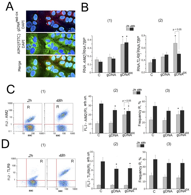
Figure 2. The exposure to gDNAOX (50 ng/mL) leads to a transient increase in expression cytoplasmic DNA sensor AIM2, while not changing expression levels of TLR9.
A - intracellular localization of AIM2 (FITC-conjugated antibodies) and labeled probe gDNAred-ox (x40). B – the ratio of the levels of AIM1 [1] and TLR9 [2] – encoding RNAs to the levels TBP-encoding reference mRNA in cells exposed to gDNA or gDNAOX for 2 hrs (grey columns) and 48 hrs (black columns).
C and D – Flow cytometry detection of AIM2 (C) and TLR9 (D) expression in MCF-7. Cells were stained with AIM2 (C) or TLR9 (D) antibody (secondary PE-conjugated antibodies). Panels D [1] and E [1] – control cells plots: FL2 versus SSC. R: gated area. Panels C [2] and D [2]: median signal intensity of FL2 (R) in MCF-7 cells (mean value for three independent experiments). Panels C [3] and D [3]: relative proportions of AIM2- or TLR9-positive cells in R gates [1]. Background fluorescence was quantified using PE-conjugated secondary antibodies.
*p < 0.05 against control group of cells, non-parametric U-test.

