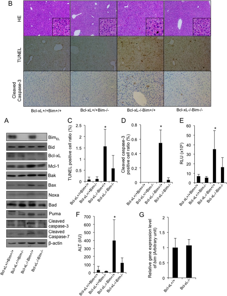
FIGURE 1.
The disruption of Bim alleviated spontaneous hepatocyte apoptosis in the absence of Bcl-xL. A–F, the offspring from the mating of bim±bcl-xflox/floxalb-cre mice with bim±bcl-xflox/flox mice were examined at 6 weeks of age. Bcl-xL+/+ and Bcl-xL−/−, bcl-xflox/flox and bcl-xflox/floxalb-cre, respectively. A, Western blot analysis of whole liver lysates for the expression of BimEL, Bid, Bcl-xL, Mcl-1, Bak, Bax, Noxa, Bad, Puma, cleaved caspase-3, cleaved caspase-7, and β-actin. B, representative images for liver histology stained with hematoxylin-eosin (HE), TUNEL, and cleaved caspase-3 (original magnifications, ×100 (large panels) and ×400 (insets)); black arrows indicate apoptotic bodies. C, TUNEL-positive cell ratio; n = 8 mice/group; *, p < 0.05 versus all. D, cleaved caspase-3-positive cell ratio; n = 3 mice/group; *, p < 0.05 versus all. E, serum caspase-3/7 activity; n = 11 mice/group; *, p < 0.05 versus all. F, serum ALT levels; n = 13 mice/group; *, p < 0.05 versus all. G, offspring from the mating of bcl-xflox/floxalb-cre mice with bcl-xflox/flox mice were examined at 6 weeks of age. Bcl-xL+/+ and Bcl-xL−/−, bcl-xflox/flox and bcl-xflox/floxalb-cre, respectively. bim mRNA levels in the whole liver tissue were determined by real-time RT-PCR; n = 6 mice/group. Error bars, S.D. RLU, relative light units; I/U, international units.

