FIGURE 6.
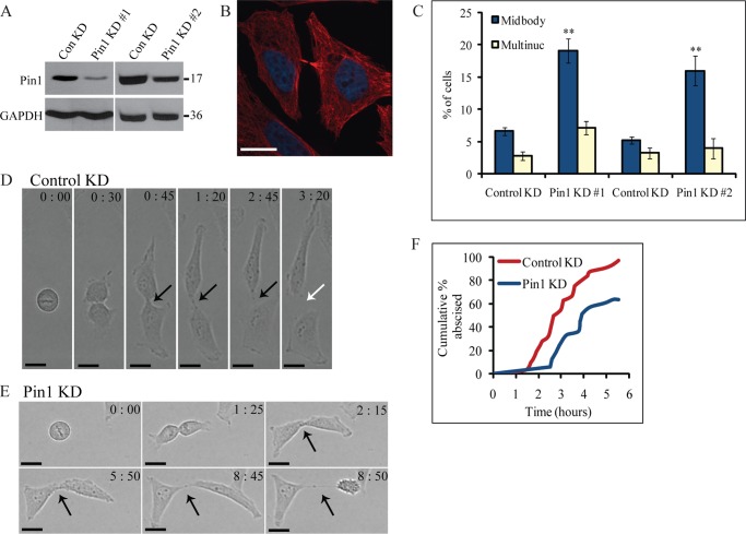
Pin1 is important for midbody abscission. A, efficiency of Pin1 depletion by siRNA. HeLa cells were treated with control siRNA or one of the two siRNAs targeting different regions of the Pin1 gene. Lysates were probed for Pin1 and GAPDH. Numbers to the right of the blots represent molecular mass standards in kilodaltons. B, representative example of cells attached by persistent midbodies following depletion of Pin1(Pin1-1 siRNA) (red, α-tubulin; blue, DNA). Scale bar represents 17 μm. C, quantification of effect of Pin1 depletion on cytokinesis. The percentage of cells exhibiting cytokinesis defects (multiple nuclei (Multinuc) or persistent midbodies) was determined upon treatment with control or Pin1 siRNA. The two Pin1 siRNAs were independently controlled so control siRNA results are shown separately. Data are represented as mean ± S.E. (n ≥ 300 cells from three or more independent experiments). Asterisks indicate differences between control and Pin1 knockdown cells; **, p < 0.005 (t test). D, division of HeLa cells after treatment with control siRNA. HeLa cells were transfected with control siRNA and randomly selected cells were followed through division by time-lapse microscopy. The time (in hours:minutes) since the beginning of DNA segregation is shown. Black arrows point to intact midbodies, whereas white arrows denote abscission. Scale bar represents 16 μm. E, Pin1 depletion causes defects in midbody abscission. HeLa cells were transfected with Pin1-1 siRNA and imaged as described in D. F, quantification of effect of Pin1 depletion on midbody abscission. The time from DNA segregation to midbody abscission was determined for each cell (n = 32 cells for control knockdown, n = 33 cells for Pin1-1 knockdown), and the cumulative percentage of cells that have abscised is plotted as a function of time. ConKD, control knockdown.

