
FIGURE 4.
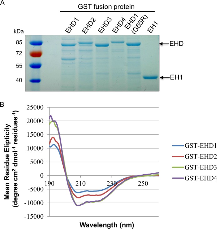
GST fusion protein purification and the secondary structure of purified EHD proteins. A, the purified GST fusion proteins were detected by SDS-PAGE and Coomassie Blue staining. B, the folding of purified EHD proteins was analyzed by circular dichroism.