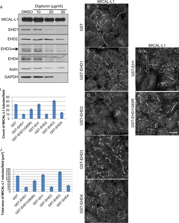
FIGURE 5.
Semipermeabilized cell system for measuring vesiculation and tubulation of TRE. A, cells were either treated with dimethyl sulfoxide (DMSO) or with 10, 20, or 30 μg/ml digitonin for 40 s. The protein levels of MICAL-L1, EHD1, EHD2, EHD3, EHD4, actin, and GAPDH were detected by immunoblotting. B–H, after permeabilization with 20 μg/ml digitonin, cells were incubated with either GST alone (B), GST-EHD1 (C), GST-EHD2 (D), GST-EHD3 (E), GST-EHD4 (F), GST-EH1 (G), or GST-EHD1G65R (H) for 30 min at 37 °C. The status of TRE (as depicted by endogenous MICAL-L1) was observed under these conditions. I and J, the number of MICAL-L1 tubules (I) and the total area of MICAL-L1 tubules (J) per field were quantified from three independent experiments using ImageJ. Error bars denote S.E. Scale bar, 10 μm.

